
+38(066)493-43-78
Показать все контакты

6) Discussion
The injections of MD-Muscle, lidocaine and sterile saline solution into the trigger point(s) of the masseter muscle(s) in the treatment to reduce myofascial pain at the same points had different results in the 3 Groups.
– The best results were obtained in Group I – MD-Muscle: better antinociceptive results (VAS reduction -4.3 = -53.75%) and reduction in EMGs values (-32.9 μV = -59.2%).
– Authors - Operational sites
Randelli F., GiaiVia A., Mazzoleni M., Brioschi M.: Centro di Chirurgia dell’Anca e Traumatologia [Hip surgery & Trauma Centre] - I.R.C.C.S Policlinico San Donato (San Donato Milanese, Milan - Italy);
Menon A., Sciancalepore F.: Azienda Socio Sanitaria Territoriale [Local Public Health Authority] Centro Specialistico Ortopedico Traumatologico Gaetano Pini-CTO [Gaetano PiniCTO Specialised Orthopaedic Trauma Centre], 1a Clinica Ortopedica [1st Orthopaedic Clinic], Milan - Italy;
Gagliano N.: Dipartimento di Scienze Biomediche per la Salute [Department of Biomedical Sciences for Health] – Università degli Studi di Milano, Milan - Italy.
1) Foreword
The tissues of the osteo-arthro-myofascial system, in particular the soft tissues and, of these, especially the joint capsules, ligaments and tendons, throughout life are subject to functional overload, accidents and ageing → inflammation → pain → degenerative evolution and consequent functional alterations.
2) Collagen - Metabolism
The physiological turnover of COL requires that its degradation by the matrix metalloproteinases (MMPs) is regulated by the tissue inhibitors of metalloproteinases (TIMPs).
– This physiological homeostatic regulation allows the body to have effective new COL at its disposal at all times, therefore biosynthesis must prevail slightly over biodegradation. When the action of the MMPs prevails over that of the TIMPs, as occurs physiologically from the age of ≈ 60 years, there is an evolution of the pathological situation towards tissue degradation. The MMPs are controlled by proinflammatory cytokines and by the ROS (reactive oxygen species), the TIMPs are regulated by anti-inflammatory cytokines (FIG. 2).
It is, once again, the good “equilibrium” of the Th1/Th2 cytotypes of the immune balance that regulates the synthesis and degradation of COL.
3) Tendons - Histology
The tendons are composed primarily by 1) type 1 COL fibres; 2) extracellular matrix rich in glycosaminoglycans (GAGs) and in proteoglycans (PGs).
– The special structure of COL provides the tendon with tensile strength, whereas the extracellular matrix provides the COL fibres with a scaffold.
Cell elements are observed between the COL fibres: the tenocytes (fibrocytes with a triangular or – more often – elongated and jagged appearance), promotors of the synthesis of matrix and pre-procollagen (FIG. 6).
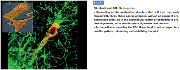
COL assembles first to form tropocollagen, which comes together to form fibrils, fibres, 1st, 2nd and 3rd order fascicles and, eventually, the tendon.
COL is synthesised inside the tenocytes and assembled outside of them, thereby making it available to the tissues.
4) Purpose of the study
In vitro analysis of the effects of MD-Tissue on human tenocytes, in order to investigate and understand the molecular mechanisms underlying the action of this Medical Device and – therefore – how it is able to affect tendon homeostasis and repair.
5) Experimental cytotype
Cultures of differentiated human tenocytes obtained from biopsy specimens of heathy tendons of the gluteus minimus muscle taken from 8 volunteer patients (4 M, 4 F; age 64.8 ± 7.2 years), undergoing hip replacement surgery.
The tenocytes were cultivated:
A) on plates containing a thin coating of MD-Tissue, or
B) on plates not containing MD-Tissue [no coating (NC) control].
Author’s notes – SUMMARY
This is the first basic (preclinical) trial on a Guna Collagen Medical Device (MD-Tissue).
– Adding MD-Tissue to a culture of human tenocytes harvested surgically from healthy tendons produces statistically significant effects on:
A) Tenocyte proliferation
B) The neosynthesis of type I collagen by inhibiting its degradation
C) Tenocyte migration for wound repair.
• These 3 experimental findings are of great importance for understanding the molecular mechanisms of repair with MD-Tissue of damaged tendons (overuse, conditions of various aetiologies, ageing).
The action of stem cells and PRP (Platelet-Rich Plasma) is akin to the action of MD-Tissue; however, they have generated conflicting results and have high (or very high) costs in addition to their operational complications.
CONCLUSIONS
Two articles have been published regarding the therapeutic use of MD-Muscle in trigger points, the first (Stanˇa, 2016-17) in the treatment of piriformis syndrome, the second (NiteckaBuchta et al., 2018), which is analytically presented in this paper, in the treatment of myofascial pain of the masseter muscle.
– Injection therapy of the myofascial trigger points is still, often and practically, the same as that proposed by Simons & Travell (1983), 35 years ago: local anaesthetic (Procaine, Lidocaine, Mepivacaine).
In addition, local injections with sterile saline solution, steroids and botulinum toxin have been, and still are, used. Local anaesthetics (amongst others, Shah et al., 2015), steroids (amongst others, Fredberg, 2007), and botulinum toxin (amongst others, Davies & Barnes, 2000), when injected locally, present potentially severe local and systemic side effects.
As regards sterile saline solution, Nitecka-Buchta et al., 2018 (in this paper) obviously consider it a neutral control, rather than a control proper.
Indeed, sterile saline solution reduces pain by just one fifth (-20%) and reduces the EMGs voltages before/after therapy (2nd and last follow-up) by just one seventh (-14%).
– Considering the critical mass of clinical studies on the efficacy of Guna Collagen MDs, true evidence of their molecular mechanisms of action was lacking.
Having demonstrated that the Guna Collagen MDs are clinically efficacious, it remained to be seen how and why they were.
The response is provided by a very recent high-quality study, the result of a partnership between 3 highly-specialised Italian centres, that was recently published in Cells (see above).
In chronic painful rheumatic/joint diseases, each cycle of therapy with Guna Collagen Medical Devices (between 6 and 10 sessions) must be repeated at least once a year, as the biological halving time of COL is 360 days at the most (Olsen, 1983).
– In the light of the great many specific and substantiated clinical findings it can now be confidently stated that Guna Collagen Medical Devices are 1) rapidly efficacious, 2) safe, 3) easy and straight-forward to apply and 4) allow good adherence from both the patient’s and the physician’s standpoint.

